Researchers at the University of New South Wales, Sydney, Australia, have reported a new study indicating that RPE curvature may provide a promising quantitative outer retinal biomarker for AMD. The researchers suggested that the “curvature” of outer retinal bands may provide a quantitative metric to correlate the measurement with AMD pathology and these measurements may be used to screen for intermediate AMD. The investigators evaluated the prognostic ability of retinal pigment epithelium (RPE) and ellipsoid zone (EZ) curvature for late AMD against outer retinal band continuity, pigmentary abnormalities, reticular pseudodrusen and drusen volume.
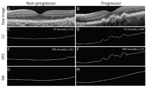
According to researchers, RPE curvature can be quantified by adapting a method for measuring river curvature—sinuosity. This RPE sinuosity was determined by calculating the ratio of RPE to Bruch’s membrane length on foveal OCT B-scans. The researchers state that the “sinuosity was found to significantly correlate with AMD lesions, including drusen, pigmentary abnormalities, and reticular pseudodrusen, and able to screen for intermediate AMD with excellent performance (area under the curve ≥0.8–0.9)”. The RPE sinuosity appeared to be robust with physiologic variations in retinal thickness, systemic disease and visual acuity and therefore the approach may be generalizable to different clinical populations. Following their research results, the researchers showed that a mean follow-up time for progressors versus non-progressors was 4.4 and 3.6 years. The RPE sinuosity was strongly associated with pigmentary abnormalities (P = 0.001) and drusen volume (P = 0.004), but not reticular pseudodrusen (RPD)(P = 0.28). RPE sinuosity >1.03 was the strongest predictor of late AMD developing within 5 years (15 [2.9–75]) and across the study period (25 [2.3-282]). Drusen volume >0.03 mm3 was the strongest predictor of progression within 2 years (33 [2.5–426]), and RPD could not independently predict progression within any time frame.
Figure 1: Sinuosity was calculated from the length of segmentation lines on foveal OCT B-scans. After extracting the raw scans (A, B), images were binarized to isolate (C, D) EZ, (E, F) RPE, and (G, H) Bruch’s membrane segmentation lines. Baseline EZ and RPE segmentation lines for an eye with stable iAMD (left column) are contrasted against an eye with iAMD that progressed to late disease within the study period (right column).
[The research work is licensed under a Creative Commons Attribution-Non Commercial-No Derivatives 4.0 International License, authored by Cheung R et al, entitled by, “Retinal pigment epithelium curvature can predict late age-related macular degeneration”. Invest Ophthalmol Vis Sci. 2024;65(12):7].
The study provides a valuable tool for the very large AMD populations in multiple countries and the researchers commented that, “this study explores sinuosity as an alternative metric for describing outer retinal integrity. Specifically, the prognostic capability of RPE and EZ sinuosity for late AMD was compared to qualitatively measured outer retinal band continuity and other current, clinical AMD biomarkers. We hypothesized that (1) both quantitative (sinuosity) and qualitative (subjective) assessments of outer retinal band integrity could predict late AMD, and (2) their performance would be noninferior to current prognostic biomarkers”.

