Researchers in Canada, have reported the first quantitative choroidal assessment in a cohort of patients with genetically characterized retinitis pigmentosa (RP). The results of the study suggest that choroidal thickness (CT) changes in RP “are not explained solely by age-related choroidal thinning, nor by SE (spherical equivalent) but seem to be dynamic and reactive to degree and rate of retinal degeneration.” The researchers indicated that a better understanding of choroidal characteristics “may identify further applications (e.g., staging, predictors of progression) of choroidal biomarkers in RP and may guide patient selection for trials and post approval treatments.”
Clinical trials use multiple metrics for psychophysical tests, electrophysiological tests, multimodal imaging and adaptive optics however, analysis of choroidal structure, with genetically characterized RP (autosomal dominant (AD), autosomal recessive (AR), and X-linked (XL) subgroups), could also provide other valuable metrics. In their current study in Vancouver, researchers used choroidal parameters (thickness, area, volume, and choroidal vascularity index (CVI)) to assess for functional relationships and predictors of degenerative pathologies. A normal central thickness of the choroid are approximately 225 to 300 μm, supporting the metabolic demands of the outer retina (see Table 1 below). Choroidal blood flow is dynamic in health and disease and these changes are quantifiable – choroidal thickness [CT], choroidal vascularity index [CVI] – as markers of disease activity. Consequently, researchers propose that choroidal anatomy may be warranted in patients with RP and other IRDs.
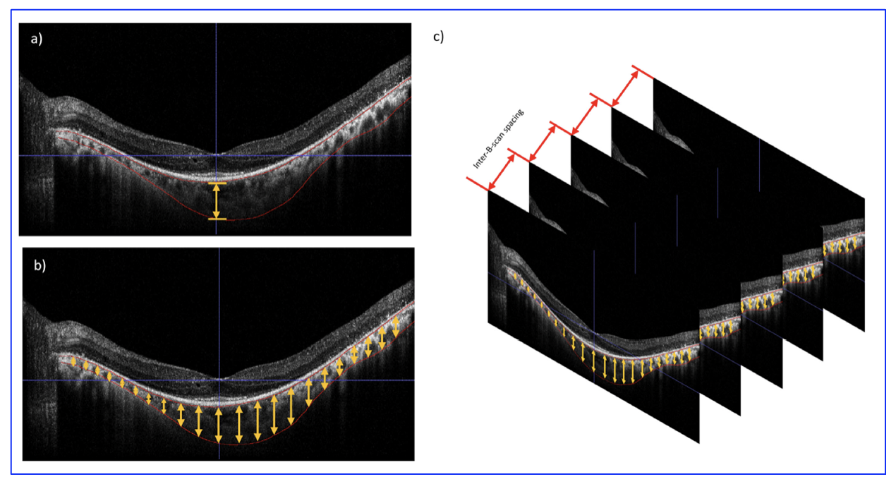
Figure 1. Technique for measuring choroidal parameters. (a) Choroidal thickness (CT), (b) choroidal area (CA), (c) choroidal volume (CV). [The research work is licensed under a Creative Commons Attribution-Non Commercial-No Derivatives 4.0 International License, cited by Stephenson, et al., entitled by: “Quantitative Choroidal Analysis of Molecularly Characterized Retinitis Pigmentosa”, Investigative Ophthalmology & Visual Science, July 2025, Vol. 66, 11, 2025; doi.org/10.1167/iovs.66.9.11].
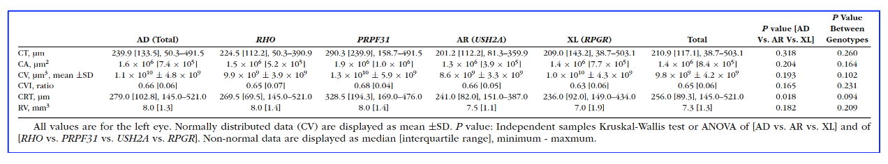
Table 1: Choroidal and Retinal Parameters for Genotype and Inheritance Groups [Stephenson, et al., entitled by: “Quantitative Choroidal Analysis of Molecularly Characterized Retinitis Pigmentosa”, Investigative Ophthalmology & Visual Science, July 2025, Vol. 66, 11, 2025; doi.org/10.1167/iovs.66.9.11].
In their analysis, a cohort of sixty-five patients (mean age, 47.3 ± 19.5 years; 52.3% female) showed that CT was thinner in RP patients than controls (P = 0.003). The proportions from each genotype group were USH2A (n = 23 [35.4%]), RPGR (n = 20 [30.8%]), RHO (n = 14 [21.5%]), and PRPF31 (n = 8 [12.3%], Table 1), providing inheritance groups of similar size. A thinner choroid was associated with older age (r = −0.512; P < 0.001) and worse BCVA (r = 0.298, P = 0.002) but not SE (P = 0.194). Although variable, no statistically significant differences were found for choroidal measures between groups. Leptochoroid (≤100 µm) was associated with advanced age (P < 0.001) and worse BCVA (P = 0.032), but not greater myopia (P = 0. 533). Greater CVI was only associated with better BCVA (P < 0.001) and no other parameters. The researchers commented that, “to our knowledge, this is the first quantitative report of choroidal features in RP which compares large groups with known genotypes. We present the study in the context of retinal structure and function.”