Researchers at Nuffield Laboratory of Ophthalmology, University of Oxford and the Oxford Eye Hospital, NHS Foundation Trust, has reported that a surrogate biomarker (comprising the ellipsoid zone (EZ) within OCT retinal images) may provide a valuable prediction tool of visual function to identify appropriate patients for enrollment in clinical trials. biomarker EZ may comprise. The study was focused on clinical trials for X-linked retinitis pigmentosa (RP) using optical coherence tomography (OCT) and function using microperimetry will be aimed to evaluate initial eligibility and endpoints. Their study aims to determine which parameters might be most sensitive in screening new patients for enrollment.
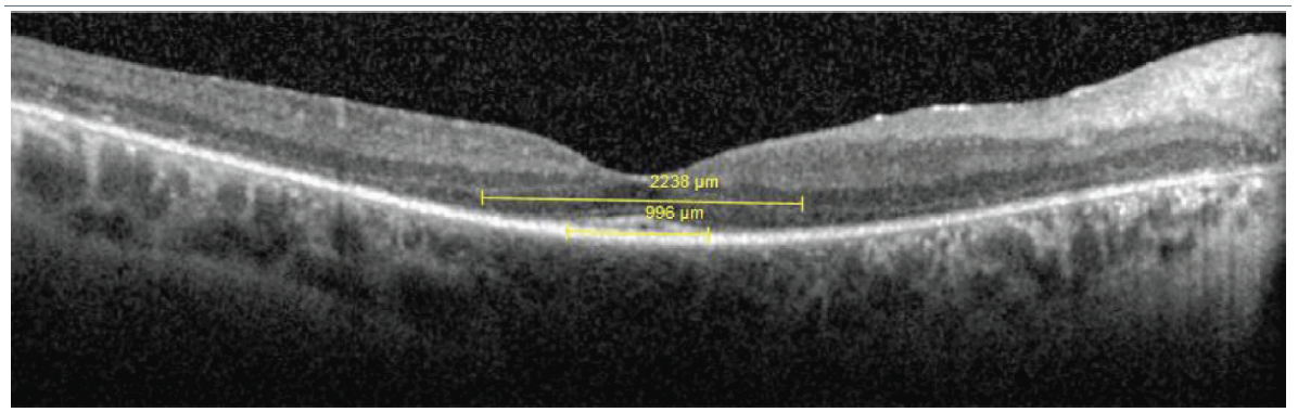
Figure 1. Representative optical coherence tomography scan indicating the remaining ellipsoid zone (EZ) and external limiting membrane (ELM). The edges of the EZ and ELM band were defined as the locations where each band met the retinal pigment epithelium, while the preserved EZ and ELM were defined as the horizontal distance between these two locations, respectively.(This work is licensed under a Creative Commons Attribution-Non Commercial-No Derivatives 4.0 International License; Christou et al, Establishing Clinical Trial Endpoints in Selecting Patients for RPGR Retinal Gene Therapy, Transl Vis Sci Technol.2024;13(9):18).
Measurement of the ellipsoid zone (EZ) and the external limiting membrane (ELM) at the inner segment/outer segment (IS/OS) junction has recently been evaluated in recent years, following technical advances on high-resolution optical coherence tomography (OCT). Multiple studies have reported that structural measurements of the EZ may correlate well with retinal function and this type of EZ measurement may act as an efficacious biomarker of progression disease, “apparently more sensitive than functional measures such as full field electroretinogram (ERG), and kinetic or static perimetry”. In terms of designing a new clinical trial, this useful EZ measurement and its’ insights into structure / function may be valuable to screen populations at the earliest stage so that the most relevant patients can be recruited to insure clinicians collect the most useful delta change for any given experimental therapy. According to the researchers, the study collected baseline measurements for retinal structure using OCT and visual function using microperimetry in patients with XLRP (RPGR-associated RP) and then “identify potential objective biomarkers that might be suitable for screening new patients that could benefit the most from genetic treatment and contribute to the designing of clinical studies to provide the best chance for successful outcomes”.
In their study, thirty-one relevant patients (62 eyes) were recruited by the Oxford Eye Hospital in a retrospective analysis measuring the remaining ellipsoid zone (EZ) and external limiting membrane (ELM) on OCT and visual function was evaluated by using 10-2 microperimetry mean sensitivity. The results showed that a linear mixed model regression analysis indicated that EZ was significantly positively correlated to ELM (P < 0.001, R² = 0.931). EZ and ELM were significantly correlated with the microperimetry sensitivity with exponential relationship (P < 0.001, R² = 0.71 and 0.72, respectively). In brief, the researchers concluded that, “our study provides valuable insights into the potential of EZ width as a biomarker for patient selection in RPGR-associated RP gene therapy trials. By elucidating the complex interplay between structural and functional retinal biomarkers, we contribute to the ongoing efforts to advance therapeutic interventions for individuals affected by inherited retinal disorders”.